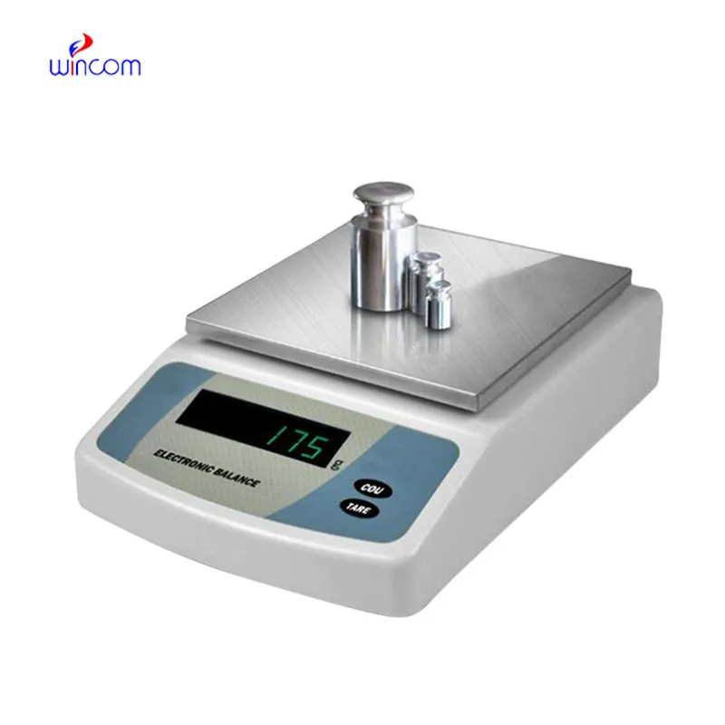
In the laboratories of hospitals and clinics, sartorius analytical balance is responsible for the precise weighing of patient samples, reagents, and pharmaceutical ingredients. Being highly precise, it minimized sample preparation errors and that is a good support for analytical results that can be reproduced. Laboratory techs apply sartorius analytical balance in the processes of quality control, method validation, and even daily operations. Reliable diagnostics, efficient laboratory workflows, and high-quality research and medical testing are the consequences of the accuracy and consistency maintained by sartorius analytical balance.

In hospital labs, sartorius analytical balance is utilized for the synthesis of analytical reagents that are employed in everyday diagnostics. Exact weighing guarantees the uniformity of the reagent concentration prior to inclusion into the automated testing systems. This application guarantees stable analytical performance in the areas of blood chemistry, immunoassays, and clinical research. By managing mass precision at the preparation stage, sartorius analytical balance allows laboratories to control reproducibility through various testing cycles, thus being an ally to reliable diagnostic results and internal quality control in hospitals.
The future of sartorius analytical balance in medical labs will put more focus on environmental stability. The advanced vibration suppression and temperature control capabilities will support precise operation even in the most crowded hospital areas. This change will make it possible to locate sartorius analytical balance nearer to the clinical workstations and this, in turn, will result in a reduction of sample transport time. Rather than moving to simpler hospital environments, sartorius analytical balance will continue to provide quick analytical preparation support and will also maintain high measurement consistency.

The maintenance of sartorius analytical balance involves the aspects of storage and inactivity care that come first. The balance should be protected from dust and vibration when it is not in active use. Periodically checking the operational status during long storage prevents unnoticed performance drift. These practices guarantee that sartorius analytical balance is still capable of accurate use in laboratories, medical and hospital settings.
In clinical labs, sartorius analytical balance serves as the primary tool for making solutions, reagents, and calibrators that are necessary for the diagnostic tests. The accuracy of weighing determines the correctness of the results of biochemical, hematological, and immunological assays. The laboratory staff depend on sartorius analytical balance for exactness, replicability, and speed. Its function secures that the diagnostic procedures of hospitals yield valid and uniform results which in turn support patient care, treatment choices, and improved laboratory quality.
Q: What is the main purpose of an Analytical Balance? A: Its purpose is mainly to measure very tiny sample masses with the utmost precision in laboratories and hospitals. Q: What is the typical weighing range of an Analytical Balance? A: The weighing range for the majority of analytical balances is from 0 up to some grams with a resolution of micrograms or milligrams. Q: What environmental controls are necessary for an Analytical Balance's operation? A: Airflow, vibration, and temperature changes should not only be avoided but also prevented in the room where the scale is situated. Q: Is an Analytical Balance permitted in a hospital laboratory? A: Yes, it has indeed found widespread usage for the preparation of reagents, calibra¬tion, and drug development applications. Q: What should be the frequency of calibration for an Analytical Balance? A: The calibration interval is subject to the degree of use and the particular laboratory requirements.
I’ve used several microscopes before, but this one stands out for its sturdy design and smooth magnification control.
The microscope delivers incredibly sharp images and precise focusing. It’s perfect for both professional lab work and educational use.
To protect the privacy of our buyers, only public service email domains like Gmail, Yahoo, and MSN will be displayed. Additionally, only a limited portion of the inquiry content will be shown.
Hello, I’m interested in your centrifuge models for laboratory use. Could you please send me more ...
I’m looking to purchase several microscopes for a research lab. Please let me know the price list ...
E-mail: [email protected]
Tel: +86-731-84176622
+86-731-84136655
Address: Rm.1507,Xinsancheng Plaza. No.58, Renmin Road(E),Changsha,Hunan,China

 af
af
 es
es
 ar
ar
 tr
tr
 sw
sw
 pt
pt
 th
th
 ur
ur
 bn
bn
 ne
ne
 vi
vi
 km
km
 lo
lo
 de
de
 ru
ru
 fi
fi
 nl
nl
 fa
fa
 fr
fr
 ko
ko