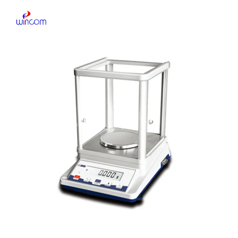
electronic price platform scale is an extremely accurate device that is specifically designed for weighing little amounts in labs and hospital pharmacies. The instrument's sensitivity implies accurate preparation of samples for diagnostic testing, reagent making, and the production of drugs. The lab personnel consider it vital to get repeatable measurements, perform calibration checks, and validate their standards using electronic price platform scale. The right use of electronic price platform scale will bring about the smoothness of clinical workflows, research activities, and quality control, thus providing accuracy and reliability to all analytical processes in hospitals and laboratories.
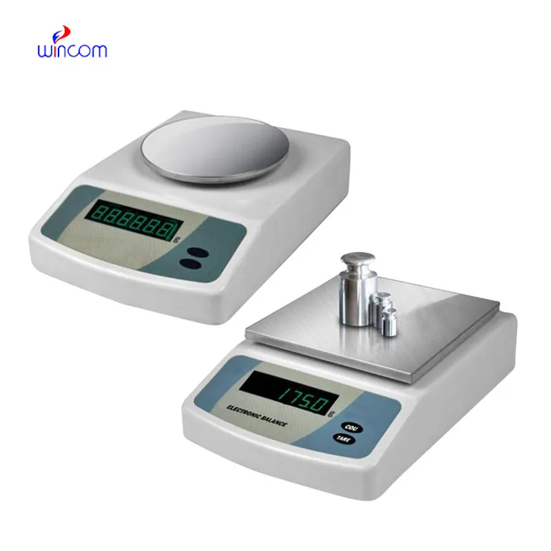
electronic price platform scale are used in clinical laboratories for making calibration references that are then checked on the analytics machines. Exact weighing guarantees that the calibration materials keep the same mass values throughout the uses they are subjected to. This application endorses instrument accuracy checks, routine audits in laboratories, and compliance with government regulations. By being a part of trustworthy calibration workflows, electronic price platform scale is a major factor and, thus, a contributor to measurement accuracy kept in hospitals' diagnostic devices.
electronic price platform scale will be an advanced generation with self-diagnostic features as the norm. Predictive monitoring of internal parts will assist laboratory teams to schedule maintenance activities in a much more efficient manner. This will lead to the continuous operation of hospital laboratories where downtime is a direct impact on both clinical workflows and research schedules.
electronic price platform scale are used in hospital settings and are good to strict contamination control measures. So, spills on the weighing chamber must always be contained. Residue buildup that may affect sensitivity is minimized by cleaning after each weighing session. The laboratory staff can identify wear early during the inspection period scheduled for downtime, and this will support long-term measurement stability across clinical applications.
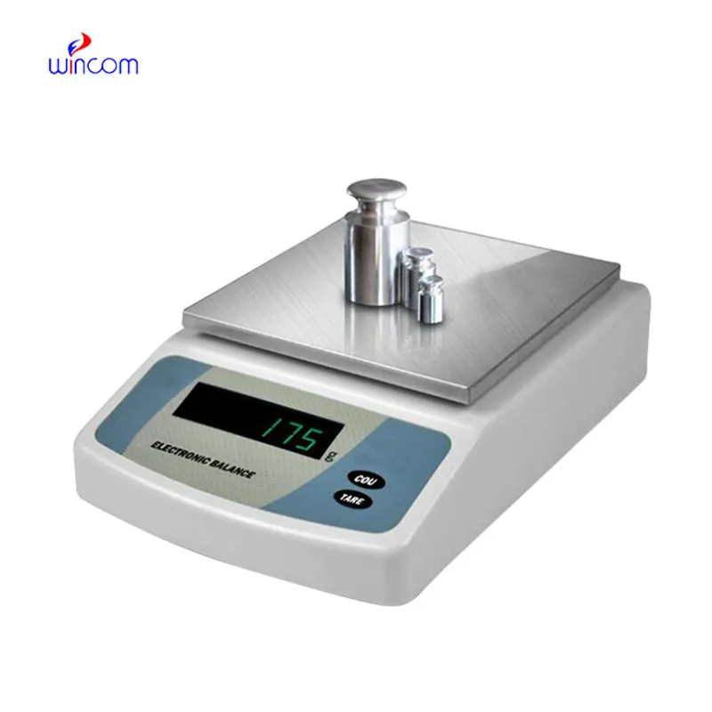
electronic price platform scale is employed in hospital labs for the reliable quality control of reagents, chemicals, and medications. Its exactness provides accurate concentrations for assays, patient treatments, and experimental protocols. The laboratory personnel regularly calibrate electronic price platform scale to rule out mistakes. Its application keeps the standard of hospital laboratories, allows the reproducibility, and builds trust in clinical and research outcomes.
Q: What is the main purpose of an Analytical Balance? A: Its purpose is mainly to measure very tiny sample masses with the utmost precision in laboratories and hospitals. Q: What is the typical weighing range of an Analytical Balance? A: The weighing range for the majority of analytical balances is from 0 up to some grams with a resolution of micrograms or milligrams. Q: What environmental controls are necessary for an Analytical Balance's operation? A: Airflow, vibration, and temperature changes should not only be avoided but also prevented in the room where the scale is situated. Q: Is an Analytical Balance permitted in a hospital laboratory? A: Yes, it has indeed found widespread usage for the preparation of reagents, calibra¬tion, and drug development applications. Q: What should be the frequency of calibration for an Analytical Balance? A: The calibration interval is subject to the degree of use and the particular laboratory requirements.
The water bath performs consistently and maintains a stable temperature even during long experiments. It’s reliable and easy to operate.
The hospital bed is well-designed and very practical. Patients find it comfortable, and nurses appreciate how simple it is to operate.
To protect the privacy of our buyers, only public service email domains like Gmail, Yahoo, and MSN will be displayed. Additionally, only a limited portion of the inquiry content will be shown.
Hello, I’m interested in your water bath for laboratory applications. Can you confirm the temperat...
We’re interested in your delivery bed for our maternity department. Please send detailed specifica...
E-mail: [email protected]
Tel: +86-731-84176622
+86-731-84136655
Address: Rm.1507,Xinsancheng Plaza. No.58, Renmin Road(E),Changsha,Hunan,China

 af
af
 es
es
 ar
ar
 tr
tr
 sw
sw
 pt
pt
 th
th
 ur
ur
 bn
bn
 ne
ne
 vi
vi
 km
km
 lo
lo
 de
de
 ru
ru
 fi
fi
 nl
nl
 fa
fa
 fr
fr
 ko
ko