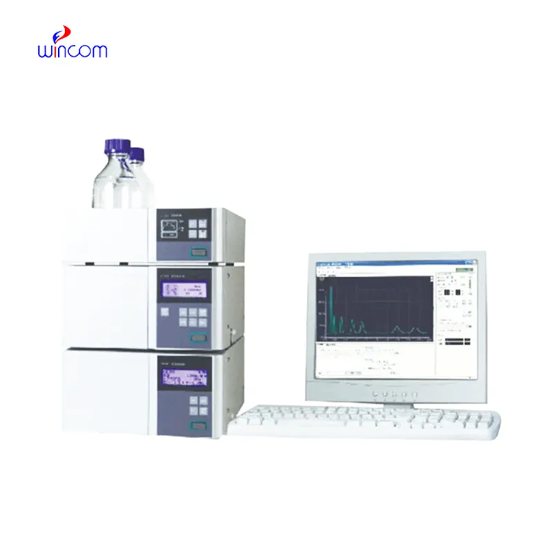
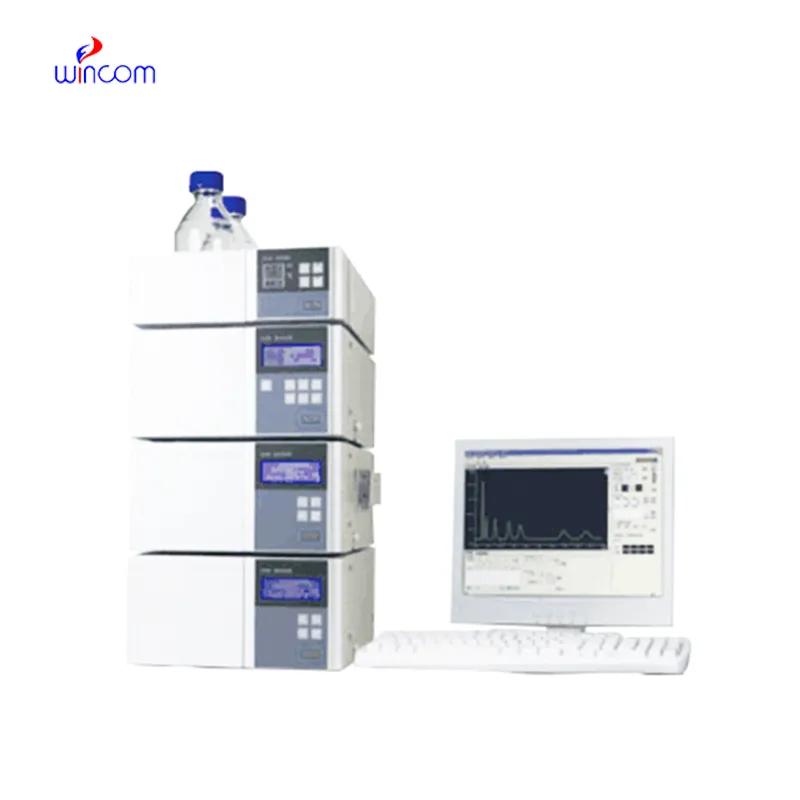
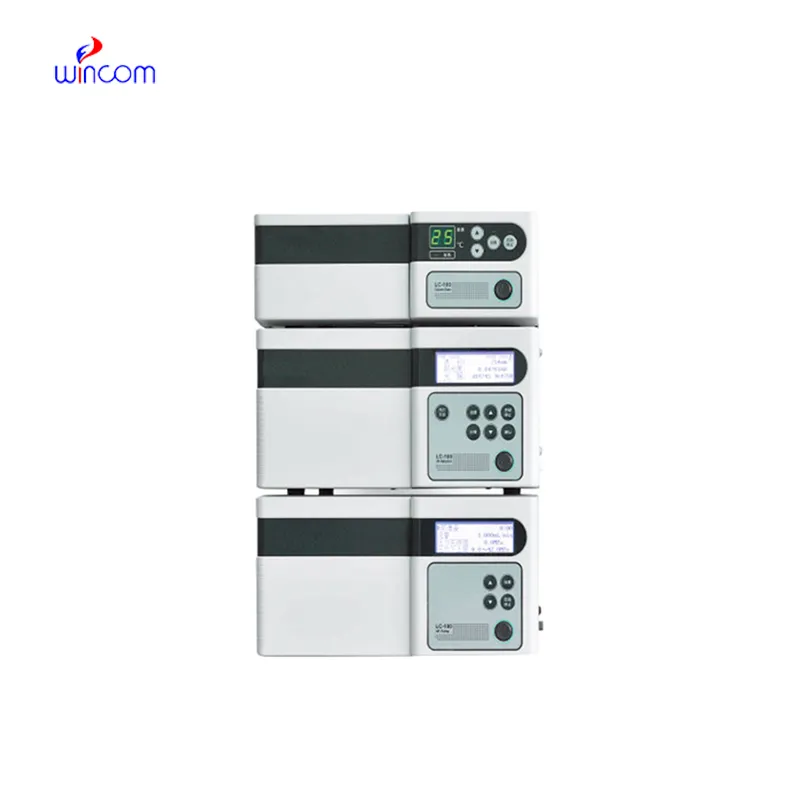
instrumentation of hplc is a primary tool in hospital and laboratory analytics. Its skills of isolating, measuring, and characterizing both chemical and biological substances enhance research as well as clinical testing. Quality control, drug testing, and testing of samples are done by laboratory technicians using instrumentation of hplc. The device's flexibility and reliability guarantee uniform performance, yielding critical analytical data that are vital for patient care, experimental validation, and smooth and fast laboratory operations in both healthcare and scientific domains.
The hospital laboratory technicians employ instrumentation of hplc to quantify the quantity of proteins or peptides. This assists in the research of biomarkers, immunotherapy studies, and analysis of responses induced by novel therapies among patients. Its accuracy and sensitivity enable the obtaining of correct results, hence aiding superior research.
The forthcoming breed of instrumentation of hplc will put a spotlight on intelligent instruments that are connected with cloud-based surveillance. Through this monitoring, hospitals will be able to gain a remote view of laboratory activities and the results of sample analysis. Lab productivity will be greatly increased by the upcoming instrumentation of hplc, and together with the new features, patient testing and therapy monitoring even in difficult clinical settings will be more accurate.
The hospital labs keep their instrumentation of hplc by adopting diligent handling and preventive maintenance. The regular examination of the columns, pumps, and connectors, along with the correct use of the solvents, aids in eliminating the problems of blockages and pressure. The lab staff is recommended to observe the cleaning and calibration according to the manufacturer's manual. The, such practices are applied, they bring about the benefits of long-term reliability, consistent separation quality, and accurate analytical outcomes in both clinical and experimental workflows.
instrumentation of hplc is a standard method in diagnostic laboratories of hospitals to keep an eye on patients’ biochemical and therapeutic figures. It quantifies drugs, hormones, and small molecules accurately. instrumentation of hplc speeds up the clinical decision-making processes of physicians and facilitates treatment modifications by supplying them with quick and precise results. It is used by hospital labs for basic patient testing, pharmacokinetic studies, and special analyses. The method’s high reproducibility makes certain that the outcomes are consistent, whereas its versatility allows for the support of many clinical applications. instrumentation of hplc has turned into an irreplaceable instrument in hospital diagnostics, which not only enhances patient management but also provides healthcare professionals with thorough molecular information.
Q: What is HPLC used for in laboratories? A: HPLC turns out to be one of the most significant and essential analytical methods in laboratories equipped with the chemical compound analysis, separation, identification, and quantification of their presence in complex samples which are the research, clinical, and pharmaceutical applications. Q: How does HPLC separate compounds? A: The HPLC separation technique is based on the different affinities of the compounds to the stationary phase and mobile phase within the chromatography column. Q: Can HPLC analyze biological samples? A: Yes, it is certainly possible to carry out analyses on various biological fluids such as blood, serum, urine, etc. for the detection of metabolites, drugs, and biomarkers. Q: How often should HPLC columns be replaced? A: The replacement of the columns must be done according to the manufacturer instructions or when the performance begins to decline, which is quite usual after heavy use or contamination. Q: What detectors can be used with HPLC? A: The analysis type determines the use of, among others, UV, fluorescence, refractive index, and mass spectrometry detectors as the common detectors.
This x-ray machine is reliable and easy to operate. Our technicians appreciate how quickly it processes scans, saving valuable time during busy patient hours.
The hospital bed is well-designed and very practical. Patients find it comfortable, and nurses appreciate how simple it is to operate.
To protect the privacy of our buyers, only public service email domains like Gmail, Yahoo, and MSN will be displayed. Additionally, only a limited portion of the inquiry content will be shown.
We’re interested in your delivery bed for our maternity department. Please send detailed specifica...
Hello, I’m interested in your water bath for laboratory applications. Can you confirm the temperat...
E-mail: [email protected]
Tel: +86-731-84176622
+86-731-84136655
Address: Rm.1507,Xinsancheng Plaza. No.58, Renmin Road(E),Changsha,Hunan,China

 af
af
 es
es
 ar
ar
 tr
tr
 sw
sw
 pt
pt
 th
th
 ur
ur
 bn
bn
 ne
ne
 vi
vi
 km
km
 lo
lo
 de
de
 ru
ru
 fi
fi
 nl
nl
 fa
fa
 fr
fr
 ko
ko