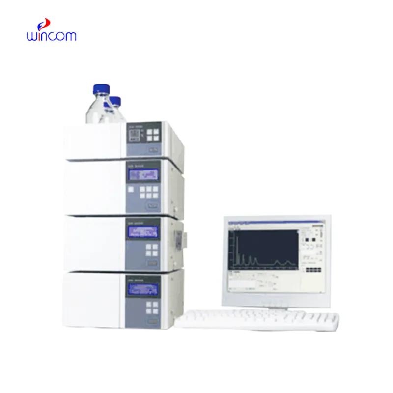
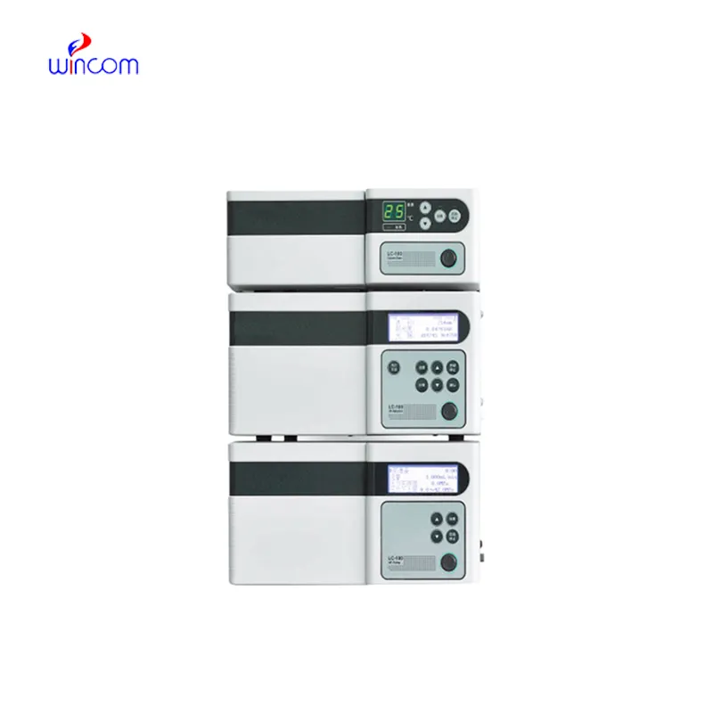
stationary phase in hplc enables labs to separate and analyze intricate mixtures with utmost precision. Through a seamless connection with current detectors, the method provides detailed profiling of both chemical and biological substances. The researchers and therapists trust stationary phase in hplc for the purposes of monitoring outcomes of experiments, method development, and cross-analyses accuracy. Its strength in dealing with various kinds of samples renders it an indispensable device in both the research and the clinical settings, thus improving reproducibility and backing up the struggling with more complex scientific and medical inquiries.
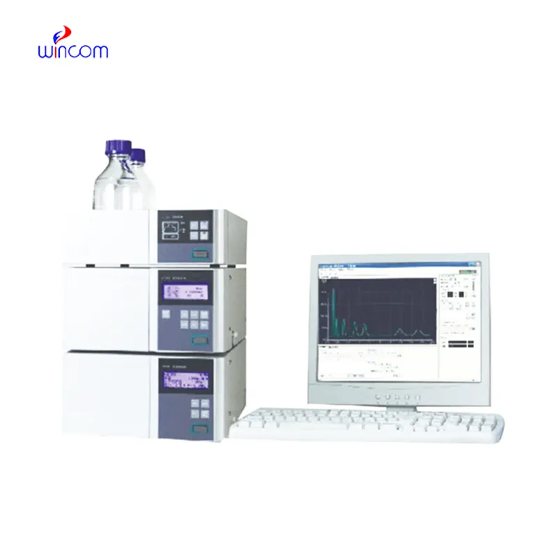
stationary phase in hplc finds extensive application in hospital laboratories for monitoring drugs therapeutically. It provides precise determination of drug levels in patients' samples, thus making safe and effective dosing possible. Metabolites are tracked, treatment progress is assessed, and unexpected drug interactions are detected by the laboratory personnel. Its high accuracy and reproducibility facilitate both medical decision-making and research, hence, stationary phase in hplc becomes an indispensable instrument in taking care of patients and analyzing the medical field.
The instruments for stationary phase in hplc of the future will be equipped with separation methods in multiple dimensions and fully automated sample preparation. The detection of trace amounts of metabolites, drugs, and biomarkers will be so accurate that hospitals and clinical laboratories will be the first to reap the benefits. The applications of stationary phase in hplc in the future will greatly help in complex diagnostics, research studies, and laboratory efficiency.
stationary phase in hplc proper care makes sure that hospital laboratories get reliable performance every time. Regular column flushing, taking care of pump and seal inspections, and using appropriate solvents are some of the measures that will keep the damage at bay and the separation efficiency high. Scheduled maintenance, system pressure control, and detector cleaning should be done by laboratory technicians to have longer instruments lifespan. Proper upkeep leads to less downtime and provides same quality analytical results in all clinical and research applications.
Therapeutic drug monitoring relies heavily on stationary phase in hplc in hospital settings. It determines the concentration of drugs in the body to guarantee efficiency and security. The laboratory staff uses it for the examination of blood, serum, or urine samples, and signifies small molecular compounds with high accuracy. By yielding consistent outcomes, stationary phase in hplc services the medics in changing the amounts and preventing side effects. Its use goes to hormone level testing, metabolite analysis, and pharmacokinetics research. With quick processing and accurate information, stationary phase in hplc is a part of the hospital patient care, making evidence-based treatment decisions possible and enhancing clinical outcomes in different departments.
Q: What types of HPLC columns are available? A: Reversed-phase, normal-phase, ion-exchange, and size-exclusion columns are the main types of columns used according to the nature of the analytes. Q: Can multiple samples be analyzed simultaneously? A: Yes, in high-throughput systems, automated sample injection and sequential analysis are among the techniques to achieve this. Q: How does temperature affect HPLC performance? A: Temperature changes can cause variations in separation efficiency and retention times; however, the majority of labs make use of precise temperature control. Q: Can HPLC be integrated with data software? A: Sure, it can be linked with laboratory software for data collection, processing, and reporting. Q: What types of laboratories use HPLC? A: HPLC is employed by hospitals, pharmaceuticals, biochemistry research, and environmental testing labs.
This ultrasound scanner has truly improved our workflow. The image resolution and portability make it a great addition to our clinic.
This x-ray machine is reliable and easy to operate. Our technicians appreciate how quickly it processes scans, saving valuable time during busy patient hours.
To protect the privacy of our buyers, only public service email domains like Gmail, Yahoo, and MSN will be displayed. Additionally, only a limited portion of the inquiry content will be shown.
Could you share the specifications and price for your hospital bed models? We’re looking for adjus...
I’d like to inquire about your x-ray machine models. Could you provide the technical datasheet, wa...
E-mail: [email protected]
Tel: +86-731-84176622
+86-731-84136655
Address: Rm.1507,Xinsancheng Plaza. No.58, Renmin Road(E),Changsha,Hunan,China

 af
af
 es
es
 ar
ar
 tr
tr
 sw
sw
 pt
pt
 th
th
 ur
ur
 bn
bn
 ne
ne
 vi
vi
 km
km
 lo
lo
 de
de
 ru
ru
 fi
fi
 nl
nl
 fa
fa
 fr
fr
 ko
ko