Through a combination of novel imaging software and the fetal dopplar, the professionals can be very precise with the details of the anatomy they have captured and even more so with the accuracy of the final result. An ergonomic design of the device contributes to the reduction of operator fatigue during long use. The device, the fetal dopplar, also comes with the ability to connect to more than one probe thus giving it the flexibility required when catering to various diagnostic needs. Besides that, the data export and connection functions of the device help to decrease the time taken in report-generating which is done through image sharing.

The fetal dopplar is a tool that medical professionals are using in different departments like pediatrics for the measurement of the size of the organs and neurology for appreciating the anatomy of the soft tissues. It makes it easy for anesthesiologists to perform nerve blocks and vascular access procedures. The fetal dopplar increase the speed and the trust of the diagnosis during both routine checkups and highly technical interventions.

The next-generation fetal dopplar solutions come with better processing capabilities and intelligent algorithms that improve the clarity of images in addition to lessening reliance on operators. The aspect of augmented reality will change the world of surgical operations. The fetal dopplar solutions will also change the face of delivering healthcare by facilitating quicker and more accurate diagnoses.

The fetal dopplar needs to be maintained properly to ensure it always provides high-quality images. The probes should never be dropped or immersed in liquid against the recommended standards. Care should be taken when handling the control board to avoid mechanical wear and tear. Updation of the firmware and testing of the fetal dopplar ensure smooth functionality during practical operations.
The fetal dopplar uses state-of-the-art ultrasound technology to deliver real-time imaging for diagnostic and monitoring purposes. It aids physicians in assessing organs, blood vessels, and soft tissue with unmatched clarity. The non-surgical device is an important tool for guiding medical procedures and making precise diagnoses. The fetal dopplar combines portability with precision, rendering it extremely useful in routine exams as well as emergency applications.
Q: How does the ultrasound scannert contribute to emergency diagnostics? A: It enables rapid assessment of internal injuries and organ conditions in time-sensitive situations. Q: Can the ultrasound scannert be upgraded with new features? A: Yes, most models support software updates to enhance performance and expand diagnostic functions. Q: What kind of power supply does the ultrasound scannert use? A: It operates on standard AC power and may include rechargeable battery options for mobile use. Q: Is the ultrasound scannert compatible with electronic medical record systems? A: Yes, it can connect to EMR systems to streamline patient data entry and storage. Q: What factors influence the image quality of the ultrasound scannert? A: Image quality depends on probe type, operator technique, and the frequency settings selected for scanning.
This ultrasound scanner has truly improved our workflow. The image resolution and portability make it a great addition to our clinic.
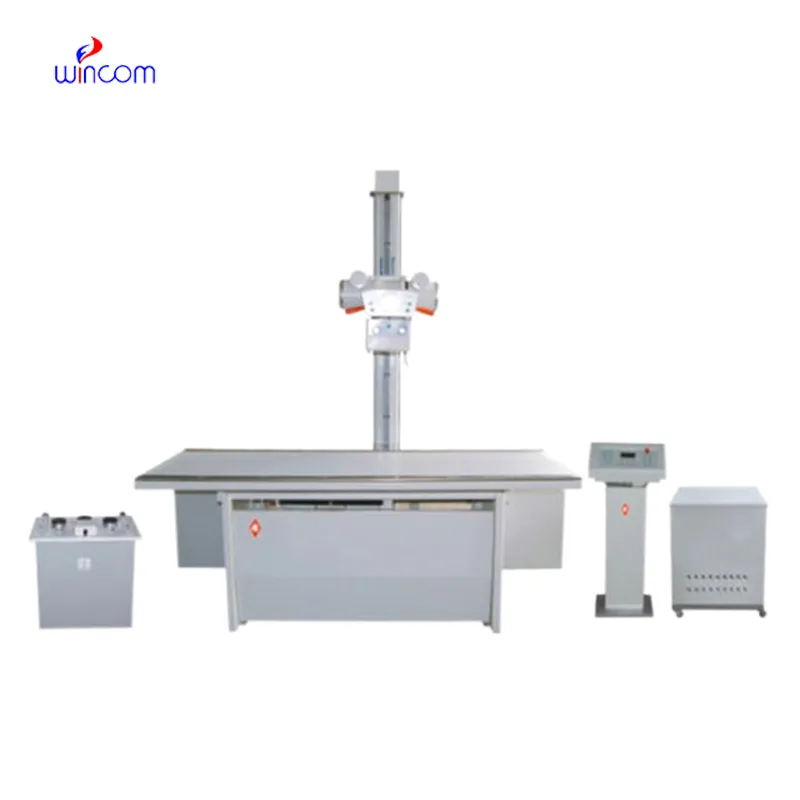
This x-ray machine is reliable and easy to operate. Our technicians appreciate how quickly it processes scans, saving valuable time during busy patient hours.
To protect the privacy of our buyers, only public service email domains like Gmail, Yahoo, and MSN will be displayed. Additionally, only a limited portion of the inquiry content will be shown.
We’re interested in your delivery bed for our maternity department. Please send detailed specifica...
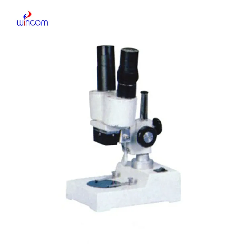
I’m looking to purchase several microscopes for a research lab. Please let me know the price list ...
E-mail: [email protected]
Tel: +86-731-84176622
+86-731-84136655
Address: Rm.1507,Xinsancheng Plaza. No.58, Renmin Road(E),Changsha,Hunan,China

 af
af
 es
es
 ar
ar
 tr
tr
 sw
sw
 pt
pt
 th
th
 ur
ur
 bn
bn
 ne
ne
 vi
vi
 km
km
 lo
lo
 de
de
 ru
ru
 fi
fi
 nl
nl
 fa
fa
 fr
fr
 ko
ko