In hospital and research facility settings, electronic analytical balance provides the critical mass measurement that is needed for delicate analyses. It is the case that reagents, samples, and medicines are weighed with the highest level of precision. Laboratory staff regard the electronic analytical balance as their helper in making the measurements, carrying out calibrations, and performing quality assurance. Besides being of great assistance in the above activities and ensuring accurate measurement in clinical diagnosis, experimental research, and drug response monitoring, electronic analytical balance also enhances overall laboratory performance and has a positive impact on the dependability of analytical results.
electronic analytical balance is used in hospital training labs for the purpose of teaching laboratory technicians and medical personnel. The learners get to do precise weighing, record the mass and use the equipment in a lab-like environment. This use boosts the formation of skills, consciousness of being accurate, and compliance with lab rules. electronic analytical balance helps in the establishment of good measurement practices hence leading to the overall betterment of lab output and uniformity in operations in the hospitals.
The future of electronic analytical balance in medical labs will put more focus on environmental stability. The advanced vibration suppression and temperature control capabilities will support precise operation even in the most crowded hospital areas. This change will make it possible to locate electronic analytical balance nearer to the clinical workstations and this, in turn, will result in a reduction of sample transport time. Rather than moving to simpler hospital environments, electronic analytical balance will continue to provide quick analytical preparation support and will also maintain high measurement consistency.
One of the main tasks in the maintenance of electronic analytical balance in the hospital laboratory is monitoring the environmental exposure. The presence of excess humidity, direct sunlight, and temperature changes should be completely ruled out. Draft shields should always be kept in a clean and working condition to cause the least possible disturbance in air during the process of weighing. These preventive activities not only help to achieve stable measurements but also aid to lessen the variability in analytical data coming from different medical testing environments.
electronic analytical balance is of great importance for developing and validating laboratory methods. One of the most important prerequisites for getting correct analytical results is the weighing of reference standards, buffers, and chemicals with absolute accuracy. Laboratory personnel depend on electronic analytical balance for constant concentration reproduction and hence, reliable testing. Its superb sensitivity plus reliable performance indeed make it a primary instrument for validating methods in the clinical, hospital, and pharmaceutical lab settings.
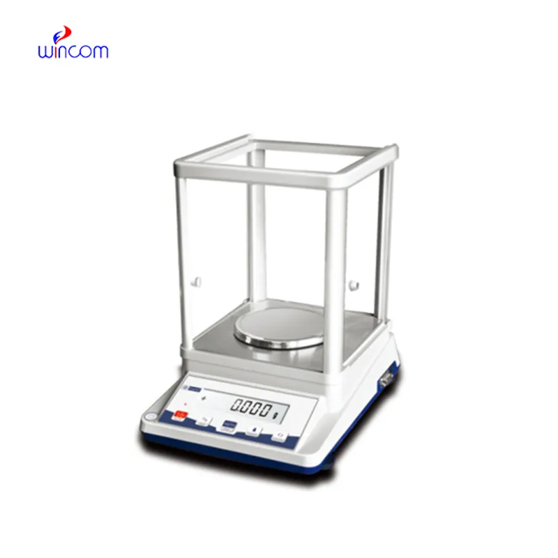
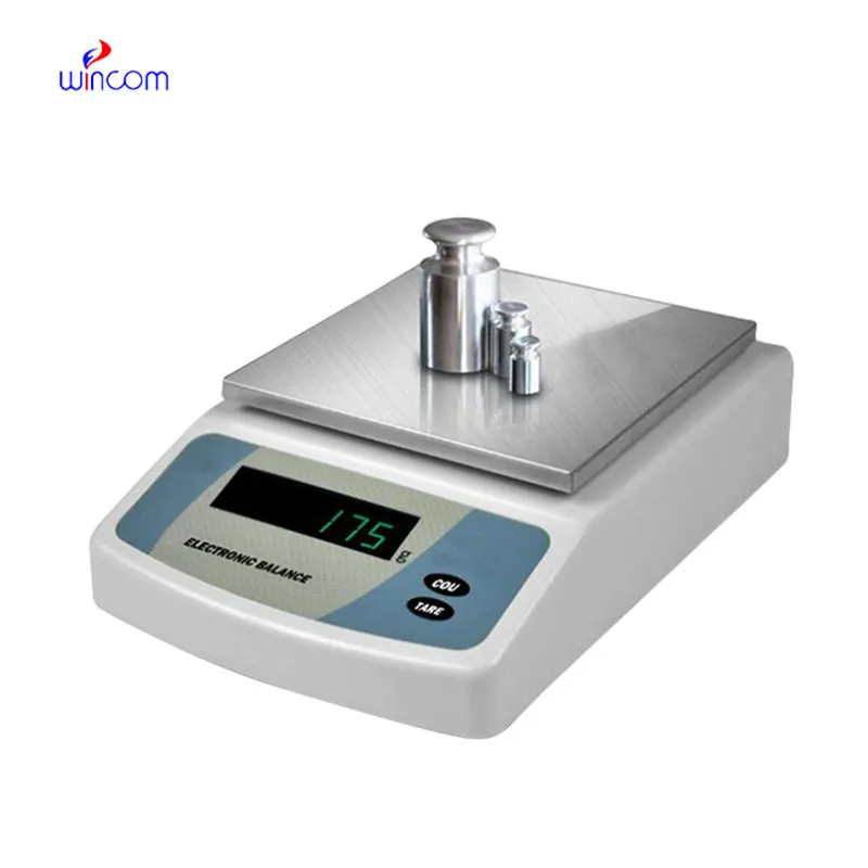
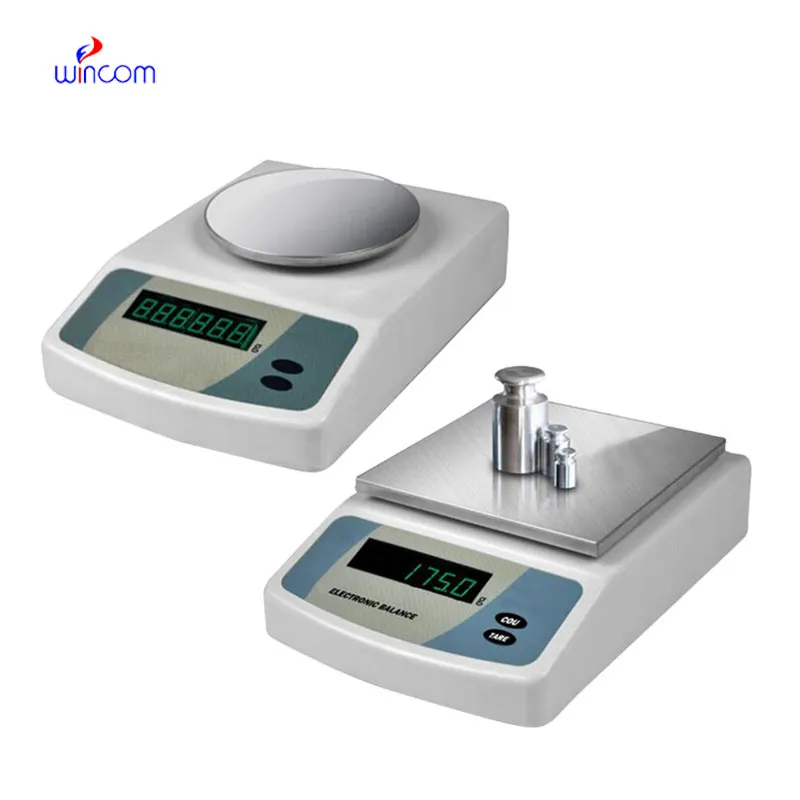
Q: What is the main purpose of an Analytical Balance? A: Its purpose is mainly to measure very tiny sample masses with the utmost precision in laboratories and hospitals. Q: What is the typical weighing range of an Analytical Balance? A: The weighing range for the majority of analytical balances is from 0 up to some grams with a resolution of micrograms or milligrams. Q: What environmental controls are necessary for an Analytical Balance's operation? A: Airflow, vibration, and temperature changes should not only be avoided but also prevented in the room where the scale is situated. Q: Is an Analytical Balance permitted in a hospital laboratory? A: Yes, it has indeed found widespread usage for the preparation of reagents, calibra¬tion, and drug development applications. Q: What should be the frequency of calibration for an Analytical Balance? A: The calibration interval is subject to the degree of use and the particular laboratory requirements.
The centrifuge operates quietly and efficiently. It’s compact but surprisingly powerful, making it perfect for daily lab use.
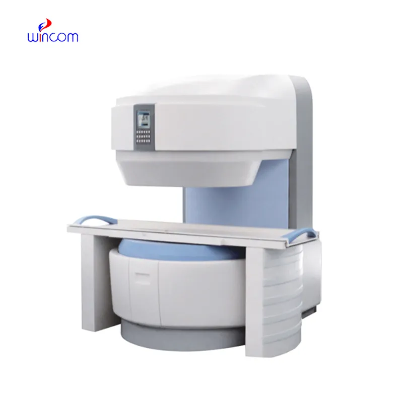
We’ve been using this mri machine for several months, and the image clarity is excellent. It’s reliable and easy for our team to operate.
To protect the privacy of our buyers, only public service email domains like Gmail, Yahoo, and MSN will be displayed. Additionally, only a limited portion of the inquiry content will be shown.
Could you please provide more information about your microscope range? I’d like to know the magnif...
We are planning to upgrade our imaging department and would like more information on your mri machin...
E-mail: [email protected]
Tel: +86-731-84176622
+86-731-84136655
Address: Rm.1507,Xinsancheng Plaza. No.58, Renmin Road(E),Changsha,Hunan,China

 af
af
 es
es
 ar
ar
 tr
tr
 sw
sw
 pt
pt
 th
th
 ur
ur
 bn
bn
 ne
ne
 vi
vi
 km
km
 lo
lo
 de
de
 ru
ru
 fi
fi
 nl
nl
 fa
fa
 fr
fr
 ko
ko