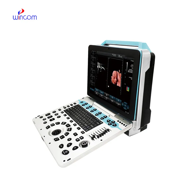
The pet ultrasound scanner, being designed for flexibility, brings the user a variety of imaging modes such as B-mode, M-mode, and color Doppler to use. Because of its small size, it can be easily moved from one place to another and that is why it can be used for examining patients in bed. The pet ultrasound scanner provides good imaging quality, which can be depended upon in cases of routine diagnostics, fieldwork, and emergency medical interventions.

In emergency departments, the pet ultrasound scanner is used for instant imaging to easily spot internal wounds and bleeding. It supports the doctor with the abdominal trauma and chest condition diagnosis. Moreover, the pet ultrasound scanner provides assistance in rural and field medical practice, delivering consistent imaging in areas with poor medical facilities.
The future of the pet ultrasound scanner may include enhancements based on artificial intelligence algorithms that can improve images and offer automatic measurements. Better transducer technologies may offer higher resolution and increased sensitivity. The pet ultrasound scanner may have an increasing role to play in personalized medicine because it may offer continuous monitoring capabilities.
The pet ultrasound scanner needs to be maintained properly to ensure it always provides high-quality images. The probes should never be dropped or immersed in liquid against the recommended standards. Care should be taken when handling the control board to avoid mechanical wear and tear. Updation of the firmware and testing of the pet ultrasound scanner ensure smooth functionality during practical operations.
The pet ultrasound scanner is more accurate in diagnostics as it captures high-resolution images of organs, tissues, and blood vessels. Design-wise flexible, it is used extensively in obstetrics, cardiology, urology, and musculoskeletal tests. Its portability and simplicity enable medical practitioners to make quick and precise evaluations. The pet ultrasound scanner makes work processes more efficient and allows for the delivery of superior patient care through real-time visualization.
Q: What is the primary function of an ultrasound scannert? A: Ultrasound scanners are designed to create real-time images of internal organs, tissues, and blood flow using high-frequency sound waves. Q: How does the ultrasound scannert ensure clear imaging results? A:It uses advanced converter technology and digital processing to enhance image clarity and contrast. Q: In what medical fields is the ultrasound scannert commonly used? A: It is widely used in obstetrics, cardiology, urology, radiology, and emergency medicine. Q: Is the ultrasound scannert safe for repeated use? A: Yes, it is non-invasive and does not emit radiation, making it safe for frequent diagnostic applications. Q: Can the ultrasound scannert store and share imaging data? A: Yes, it supports data storage, retrieval, and digital transfer for easy integration with hospital systems.
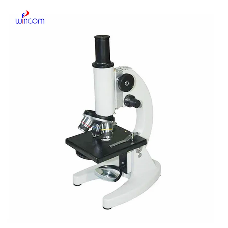
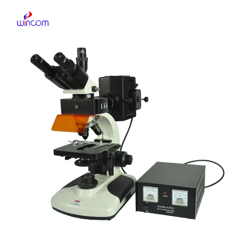
I’ve used several microscopes before, but this one stands out for its sturdy design and smooth magnification control.
This ultrasound scanner has truly improved our workflow. The image resolution and portability make it a great addition to our clinic.
To protect the privacy of our buyers, only public service email domains like Gmail, Yahoo, and MSN will be displayed. Additionally, only a limited portion of the inquiry content will be shown.
We’re interested in your delivery bed for our maternity department. Please send detailed specifica...
We are planning to upgrade our imaging department and would like more information on your mri machin...
E-mail: [email protected]
Tel: +86-731-84176622
+86-731-84136655
Address: Rm.1507,Xinsancheng Plaza. No.58, Renmin Road(E),Changsha,Hunan,China

 af
af
 es
es
 ar
ar
 tr
tr
 sw
sw
 pt
pt
 th
th
 ur
ur
 bn
bn
 ne
ne
 vi
vi
 km
km
 lo
lo
 de
de
 ru
ru
 fi
fi
 nl
nl
 fa
fa
 fr
fr
 ko
ko