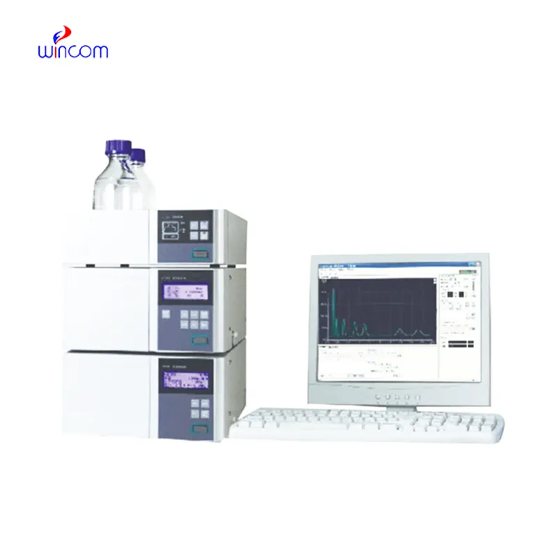
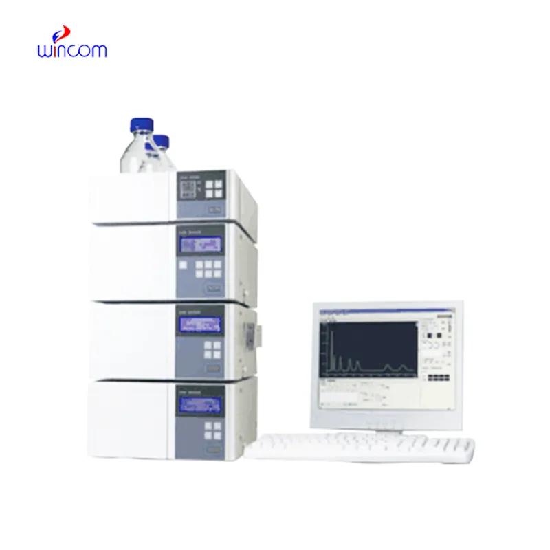
monolithic hplc column enables labs to separate and analyze intricate mixtures with utmost precision. Through a seamless connection with current detectors, the method provides detailed profiling of both chemical and biological substances. The researchers and therapists trust monolithic hplc column for the purposes of monitoring outcomes of experiments, method development, and cross-analyses accuracy. Its strength in dealing with various kinds of samples renders it an indispensable device in both the research and the clinical settings, thus improving reproducibility and backing up the struggling with more complex scientific and medical inquiries.
Hospital laboratories depend on monolithic hplc column for identifying minute quantities of pharmaceuticals and therapeutic agents in difficult-to-analyze biological samples. Its use spans drug compliance testing, pharmacokinetics profiling, and tracking medications after surgery. The laboratory personnel can rely on it for exact measurement, thus increasing the efficiency of clinical treatment.
In monolithic hplc column, the evolution is probably going to be through miniaturization and portability monolithic hplc column is the main feature of the future hospital and laboratory. These advancements will let bedside or point-of-care analysis, thus, improving hospital diagnostics and reducing turnaround times. The future highlights quickness, highly reproducible measurements, and still good accuracy in patient monitoring and laboratory research.
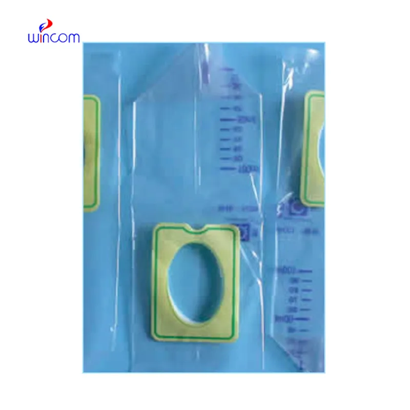
Proper handling and care of monolithic hplc column ensure continuous accuracy in the medical laboratory workflows. Cleaning of flow paths, checking detector response, and verifying pump performance are the essential maintenance tasks. Along with the column storage, solvent selection, and routine calibration, laboratory personnel must adhere to the manufacturer guidelines. Proper care enhances reproducibility, reduces downtime, and supports the consistent performance of the laboratory in hospitals and clinical research facilities.
monolithic hplc column is equipped with an in-depth examination of biomolecules like proteins, peptides, and nucleic acids. Reversed-phase, ion-exchange, and size-exclusion chromatography methods qualify scientists to get insight into the molecular properties with utmost accuracy. The application of monolithic hplc column in metabolomics studies, enzyme kinetics, and protein characterization helps in high accuracy and reproducibility. The high sensitivity level helps to detect low-molecular-weight molecules in detail and get insight into biological samples at a high level. One of the prime reasons why scientists are interested in monolithic hplc column is its ability to generate information that advances understanding at an advanced biochemistry level.
Q: What is HPLC used for in laboratories? A: HPLC turns out to be one of the most significant and essential analytical methods in laboratories equipped with the chemical compound analysis, separation, identification, and quantification of their presence in complex samples which are the research, clinical, and pharmaceutical applications. Q: How does HPLC separate compounds? A: The HPLC separation technique is based on the different affinities of the compounds to the stationary phase and mobile phase within the chromatography column. Q: Can HPLC analyze biological samples? A: Yes, it is certainly possible to carry out analyses on various biological fluids such as blood, serum, urine, etc. for the detection of metabolites, drugs, and biomarkers. Q: How often should HPLC columns be replaced? A: The replacement of the columns must be done according to the manufacturer instructions or when the performance begins to decline, which is quite usual after heavy use or contamination. Q: What detectors can be used with HPLC? A: The analysis type determines the use of, among others, UV, fluorescence, refractive index, and mass spectrometry detectors as the common detectors.
The microscope delivers incredibly sharp images and precise focusing. It’s perfect for both professional lab work and educational use.
We’ve been using this mri machine for several months, and the image clarity is excellent. It’s reliable and easy for our team to operate.
To protect the privacy of our buyers, only public service email domains like Gmail, Yahoo, and MSN will be displayed. Additionally, only a limited portion of the inquiry content will be shown.
We are planning to upgrade our imaging department and would like more information on your mri machin...
I’d like to inquire about your x-ray machine models. Could you provide the technical datasheet, wa...
E-mail: [email protected]
Tel: +86-731-84176622
+86-731-84136655
Address: Rm.1507,Xinsancheng Plaza. No.58, Renmin Road(E),Changsha,Hunan,China

 af
af
 es
es
 ar
ar
 tr
tr
 sw
sw
 pt
pt
 th
th
 ur
ur
 bn
bn
 ne
ne
 vi
vi
 km
km
 lo
lo
 de
de
 ru
ru
 fi
fi
 nl
nl
 fa
fa
 fr
fr
 ko
ko