The microscope for observing microscopic plant cells in school labs is engineered to deliver consistent performance at all magnification levels. With precision focusing knobs and a rugged mechanical stage, it offers accurate sample positioning and smooth handling. The illumination system provides even lighting for clear observation of opaque and transparent specimens. Most microscope for observing microscopic plant cells in school labs models have modular configurations, which can be customized for particular fields like biology, metallurgy, or semiconductor inspection.
The microscope for observing microscopic plant cells in school labs is critical to science and manufacturing advancement. In the medical research arena, the microscope for observing microscopic plant cells in school labs aids microscopic blood and tissue testing for accurate diagnostics. Research institutions use the microscope for observing microscopic plant cells in school labs in cell culture analysis, detecting bacterial growth, and research on biofilms. Industrial laboratory environments utilize the microscope for observing microscopic plant cells in school labs for product quality assurance and surface finishes evaluation. The microscope for observing microscopic plant cells in school labs is also applied in environmental science to support monitoring of plankton populations and particles of pollutants, to enhance ecological studies and sustainability science.

The microscope for observing microscopic plant cells in school labs will emerge hand in hand with revolutionary breakthroughs in computer science and optics. Future designs will incorporate ultra-sensitive detectors that can measure nanoscale motion in real-time. Through AI-aided enhancement, the microscope for observing microscopic plant cells in school labs will facilitate predictive medicine and materials science analysis. Enhanced portability will allow researchers to employ small units on-site or at remote sites. As further technology emerges, the microscope for observing microscopic plant cells in school labs will provide a critical portal for microanalysis and worldwide science networks.

Maintenance of the microscope for observing microscopic plant cells in school labs involves regular cleaning and preventive inspection. Always start by making sure all lenses and eyepieces are clean of dust before observing. Avoid subjecting the microscope for observing microscopic plant cells in school labs to extreme temperatures or humidity levels. Clean immersion lenses after each session and remove all the slides from the stage. Keep the microscope for observing microscopic plant cells in school labs covered when not in use to protect it from contaminants. Engage professional maintenance every year to inspect optical alignment and ensure there is smooth mechanical running.
With a microscope for observing microscopic plant cells in school labs, human man can explore the microcosm with unprecedented clarity. The instrument magnifies small samples so that exact study can be conducted in laboratories, clinics, and schools. The microscope for observing microscopic plant cells in school labs recognizes cell morphology, bacterial cultures, and intricate material surfaces. Although optical and electronic technology has been enhanced, the microscope for observing microscopic plant cells in school labs of today's time offers more magnification, image stability, and integration into digital media for efficient data registration and perception.
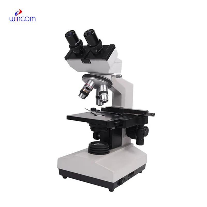
Q: What are the main parts of a microscope? A: The key components include the eyepiece, objective lenses, stage, focusing knobs, and illumination system, all working together to magnify and clarify specimens. Q: How do you clean the lenses of a microscope? A: Lenses should be cleaned using soft lens paper or microfiber cloth with a small amount of lens cleaner to avoid scratching or damaging optical coatings. Q: What magnification levels can a microscope achieve? A: Depending on the model, a microscope can typically achieve magnifications ranging from 40x to over 1000x for detailed observation of microscopic structures. Q: Why is light adjustment important in a microscope? A: Proper light adjustment ensures accurate contrast and brightness, allowing clear observation without distortion or glare during viewing. Q: Can a microscope be used for educational purposes? A: Yes, microscopes are widely used in classrooms and laboratories to teach students about biology, materials science, and microscopic analysis.
The microscope delivers incredibly sharp images and precise focusing. It’s perfect for both professional lab work and educational use.
I’ve used several microscopes before, but this one stands out for its sturdy design and smooth magnification control.
To protect the privacy of our buyers, only public service email domains like Gmail, Yahoo, and MSN will be displayed. Additionally, only a limited portion of the inquiry content will be shown.
We’re interested in your delivery bed for our maternity department. Please send detailed specifica...
I’m looking to purchase several microscopes for a research lab. Please let me know the price list ...
E-mail: [email protected]
Tel: +86-731-84176622
+86-731-84136655
Address: Rm.1507,Xinsancheng Plaza. No.58, Renmin Road(E),Changsha,Hunan,China

 af
af
 es
es
 ar
ar
 tr
tr
 sw
sw
 pt
pt
 th
th
 ur
ur
 bn
bn
 ne
ne
 vi
vi
 km
km
 lo
lo
 de
de
 ru
ru
 fi
fi
 nl
nl
 fa
fa
 fr
fr
 ko
ko