Every feature of the microscope for algae observation in environmental studies is built to provide maximum viewing accuracy and comfort to the user. The coarse and fine focus of the microscope provide control to observe large and small specimens. The microscope for algae observation in environmental studies has enhanced illumination technology to give balanced light conditions to highlight color contrasts and fine details. It has also got compatibility with digital imaging software for analysis and documentation to allow researchers to store and compare results efficiently.
Versatile in use, the microscope for algae observation in environmental studies has extensive usage in laboratories, universities, and manufacturing. It is used to provide precise observation of living organisms, minerals, and artificial materials. In life science research, the microscope for algae observation in environmental studies helps examine cellular processes and structures of genes. Metallurgists make use of it to examine grain boundaries and fatigue cracks, while chemists make use of it to examine crystalline compounds. It is also used in the textile industry to assess fiber quality and compositional structure at high magnification.

The next generation of the microscope for algae observation in environmental studies will merge optics with digital intelligence. Artificial intelligence and machine learning algorithms will interpret complex images automatically, allowing scientists to identify microscopic structures faster. Improved ergonomic interfaces that are more human-friendly and voice-controlled interfaces will improve the interaction with the microscope for the users. The microscope for algae observation in environmental studies will also be equipped with environmental sensors to provide stability and precision in the functioning. With the integration of virtual reality, scientists are now able to explore micro-worlds in interactive three-dimensional environments, expanding visual research boundaries.

To continue functioning optimally, the microscope for algae observation in environmental studies must be treated to regular maintenance with attention to detail. Clean lenses with soft strokes using microfiber cloths or dedicated wipes. Avoid spraying cleaners directly on the optics. Keep the stage and focus assembly residue and corrosion free. Always shut down when cleaning electrical components. When storing, cover the microscope for algae observation in environmental studies and place it in a dry, temperature-controlled environment. Periodic service inspections will ensure accurate focusing, smooth operation, and long-term durability.
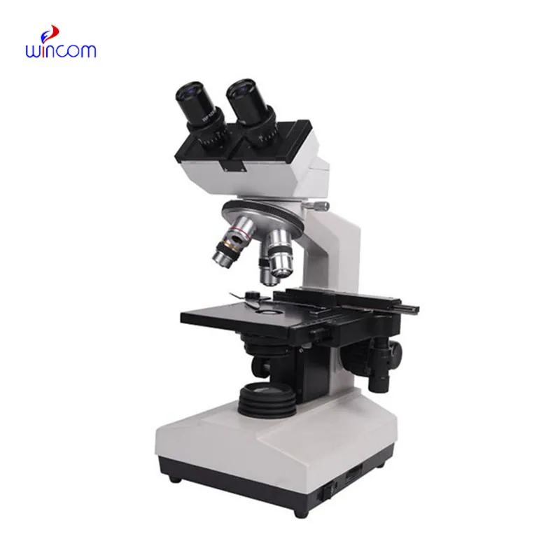
A microscope for algae observation in environmental studies is a convenient tool that magnifies microscopic materials that are invisible to the naked eye. It allows researchers, scientists, and students to view cells, microorganisms, and sensitive materials with careful attention at microscopic sizes. Modern microscope for algae observation in environmental studies models combine optical precision with electronic technology to give high-definition images and fine focusing. They are widely applied in biology, medicine, and material sciences for research, study, and instruction. With high-performance lenses and illumination systems, a microscope for algae observation in environmental studies enhances visualization to enable users to examine texture, shape, and structure at the microscopic level with utmost clarity and accuracy.
Q: What are the main parts of a microscope? A: The key components include the eyepiece, objective lenses, stage, focusing knobs, and illumination system, all working together to magnify and clarify specimens. Q: How do you clean the lenses of a microscope? A: Lenses should be cleaned using soft lens paper or microfiber cloth with a small amount of lens cleaner to avoid scratching or damaging optical coatings. Q: What magnification levels can a microscope achieve? A: Depending on the model, a microscope can typically achieve magnifications ranging from 40x to over 1000x for detailed observation of microscopic structures. Q: Why is light adjustment important in a microscope? A: Proper light adjustment ensures accurate contrast and brightness, allowing clear observation without distortion or glare during viewing. Q: Can a microscope be used for educational purposes? A: Yes, microscopes are widely used in classrooms and laboratories to teach students about biology, materials science, and microscopic analysis.
The delivery bed is well-designed and reliable. Our staff finds it simple to operate, and patients feel comfortable using it.
The microscope delivers incredibly sharp images and precise focusing. It’s perfect for both professional lab work and educational use.
To protect the privacy of our buyers, only public service email domains like Gmail, Yahoo, and MSN will be displayed. Additionally, only a limited portion of the inquiry content will be shown.
Could you please provide more information about your microscope range? I’d like to know the magnif...
We’re currently sourcing an ultrasound scanner for hospital use. Please send product specification...
E-mail: [email protected]
Tel: +86-731-84176622
+86-731-84136655
Address: Rm.1507,Xinsancheng Plaza. No.58, Renmin Road(E),Changsha,Hunan,China

 af
af
 es
es
 ar
ar
 tr
tr
 sw
sw
 pt
pt
 th
th
 ur
ur
 bn
bn
 ne
ne
 vi
vi
 km
km
 lo
lo
 de
de
 ru
ru
 fi
fi
 nl
nl
 fa
fa
 fr
fr
 ko
ko