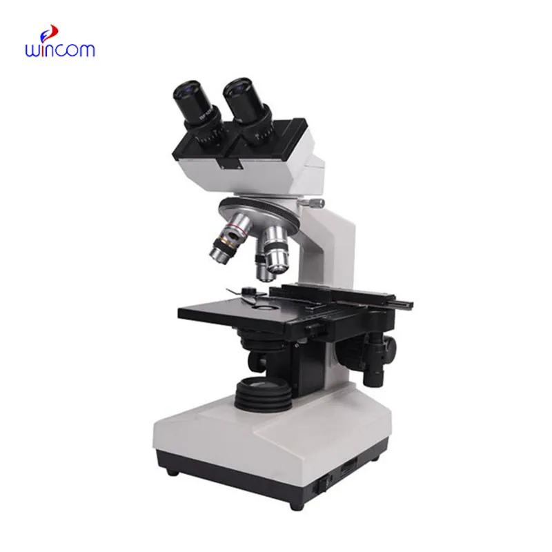
The high magnification microscope for detecting foodborne pathogens is engineered for precision and versatility, featuring adjustable magnification levels and ergonomic design for continuous use. Its optical system delivers uniform brightness and sharp focus on different specimens. Equipped with illumination controls within, the high magnification microscope for detecting foodborne pathogens maximizes contrast and clarity, enabling easier observation of delicate structures. Digital cameras and connectivity options for real-time image acquisition and sharing are included in most models. The high magnification microscope for detecting foodborne pathogens is built with durable materials to maintain stability of performance despite routine laboratory use.
The high magnification microscope for detecting foodborne pathogens is today a vital component of sectors that involve precise observation. In schools, it promotes scientific investigation by allowing students to observe living cells and microscopic organisms. Biomedical professionals apply the high magnification microscope for detecting foodborne pathogens in diagnostic testing of tissues and histological analysis. The high magnification microscope for detecting foodborne pathogens is also applied in industrial testing, where engineers study welds, coatings, and fractures. The high magnification microscope for detecting foodborne pathogens assists environmental scientists in studying microorganisms in soil and water environments, ensuring sustainability and preservation.
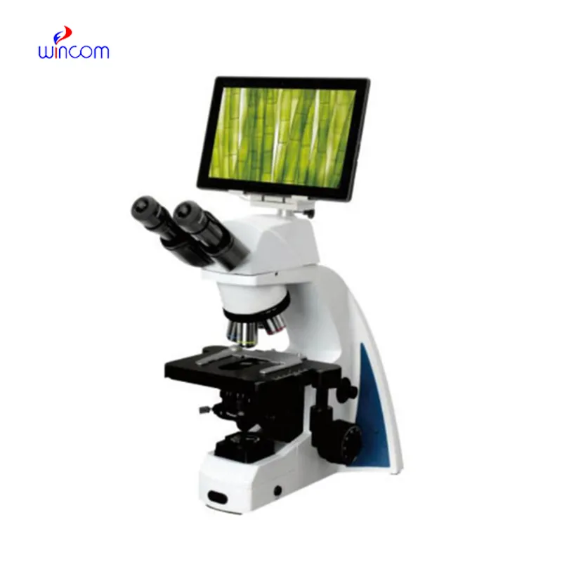
With the progress of technology, the high magnification microscope for detecting foodborne pathogens will turn into a smarter and more interactive research tool. Compatibility with AI will allow it to detect patterns, recognize anomalies, and measure data automatically. The high magnification microscope for detecting foodborne pathogens will also make remote diagnostics possible, where the samples from every corner of the world can be diagnosed remotely by specialists. Advances in imaging sensors and optical systems will provide better depth resolution and faster capture rates. These will expand the uses of the high magnification microscope for detecting foodborne pathogens in medicine, nanotechnology, and education.

In the interest of precision and reliability, the high magnification microscope for detecting foodborne pathogens should be constantly exposed to cleanliness and maintenance. Switch it off at all times before adjusting or cleaning parts. The lenses may be cleaned with alcohol-free cleaners lightly to avoid scratching. Rotary components such as knobs and stage mechanisms value light lubrication at regular intervals. The high magnification microscope for detecting foodborne pathogens must be stored away from direct sunlight and vibration. Professional checking once a year ensures optical alignment is not affected and prevents wear from invisible damage.
The high magnification microscope for detecting foodborne pathogens allows researchers to study the world at a microscopic level with stunning detail. Using high-tech optical or electron systems, the high magnification microscope for detecting foodborne pathogens magnifies samples to reveal texture, layers, and details that are imperceptible to the human eye. From life sciences to factory quality control, uses span the range. Portable and compact models now combine ergonomic design and digital controls to offer comfort, accuracy, and dependability for extended observation periods.
Q: What is the lifespan of a microscope? A: With proper care and maintenance, a microscope can last for many years, providing consistent optical performance and stability. Q: How does the objective lens affect image quality in a microscope? A: The objective lens determines magnification and resolution; high-quality lenses produce sharper, more accurate images of specimens. Q: Can a microscope be used to view live specimens? A: Yes, many microscope models support live-cell observation, allowing users to study biological processes in real time under controlled conditions. Q: What is the function of the condenser in a microscope? A: The condenser focuses light onto the specimen, enhancing illumination and improving contrast for clear image viewing. Q: How should a microscope be transported safely? A: Carry the microscope with both hands—one under the base and one on the arm—to prevent damage or misalignment of delicate parts.
I’ve used several microscopes before, but this one stands out for its sturdy design and smooth magnification control.
The microscope delivers incredibly sharp images and precise focusing. It’s perfect for both professional lab work and educational use.
To protect the privacy of our buyers, only public service email domains like Gmail, Yahoo, and MSN will be displayed. Additionally, only a limited portion of the inquiry content will be shown.
We’re interested in your delivery bed for our maternity department. Please send detailed specifica...
Could you please provide more information about your microscope range? I’d like to know the magnif...
E-mail: [email protected]
Tel: +86-731-84176622
+86-731-84136655
Address: Rm.1507,Xinsancheng Plaza. No.58, Renmin Road(E),Changsha,Hunan,China

 af
af
 es
es
 ar
ar
 tr
tr
 sw
sw
 pt
pt
 th
th
 ur
ur
 bn
bn
 ne
ne
 vi
vi
 km
km
 lo
lo
 de
de
 ru
ru
 fi
fi
 nl
nl
 fa
fa
 fr
fr
 ko
ko