Small samples for accurate mass measurement depend on the laboratory's electronic benefit transfer delaware balance that determine the efficiency and reliability. In hospitals, it guides researchers and pharmacy staff in making medicines, calibrators, and experimental solutions. electronic benefit transfer delaware balance guarantees very low weight measurement variation which is a precondition for reproducible laboratory results. Its strength, sensitivity, and accuracy are the reasons it is indispensable in clinical laboratories, pharmaceutical testing, and biomedical research where precision is critical.

electronic benefit transfer delaware balance are used in clinical laboratories for making calibration references that are then checked on the analytics machines. Exact weighing guarantees that the calibration materials keep the same mass values throughout the uses they are subjected to. This application endorses instrument accuracy checks, routine audits in laboratories, and compliance with government regulations. By being a part of trustworthy calibration workflows, electronic benefit transfer delaware balance is a major factor and, thus, a contributor to measurement accuracy kept in hospitals' diagnostic devices.
electronic benefit transfer delaware balance will envision a new era where the user-centered interfaces of hospital workers take greater priority. Such friendly visualizations, accompanied by the directing of workflows and notifications, will prove to be helpful during the working of professionals with machines. This change will cut down the duration of training and will also bring in better quality of work in clinical laboratories. electronic benefit transfer delaware balance will not give up on the accuracy aspect while still allowing the ease of use in the hospital environments that are changing fast.
Most care routines for electronic benefit transfer delaware balance consist of planned startup and shutdown methods. Giving enough time for the warm-up process to take place guarantees that internal electronics will be operating at stable conditions. Power cuts are not allowed and this way sensitive circuits are protected. The critics of the hospitals that have implemented standard operating procedures operate daily lab analyses with a consistent measure of accuracy and at the same time prolong the life of the electronic benefit transfer delaware balance.
In clinical labs, electronic benefit transfer delaware balance serves as the primary tool for making solutions, reagents, and calibrators that are necessary for the diagnostic tests. The accuracy of weighing determines the correctness of the results of biochemical, hematological, and immunological assays. The laboratory staff depend on electronic benefit transfer delaware balance for exactness, replicability, and speed. Its function secures that the diagnostic procedures of hospitals yield valid and uniform results which in turn support patient care, treatment choices, and improved laboratory quality.
Q: What industries are the widespread users of Analytical Balances? A: Their primary application lies in laboratories, hospitals, pharmaceutical facilities, and research institutions. Q: Is it possible to measure liquids with an Analytical Balance? A: Liquids can be indirectly measured using appropriate containers. Q: What does it mean by repeatability in an Analytical Balance? A: It is the capability to provide constant results when tested under the same conditions. Q: Is it necessary for the installation of Analytical Balances to be in controlled environments? A: Controlled environments are beneficial for keeping accuracy and stability long term. Q: What is the average lifespan of an Analytical Balance? A: If taken care of and maintained properly, it will be a reliable and many years-long operating device.
The microscope delivers incredibly sharp images and precise focusing. It’s perfect for both professional lab work and educational use.
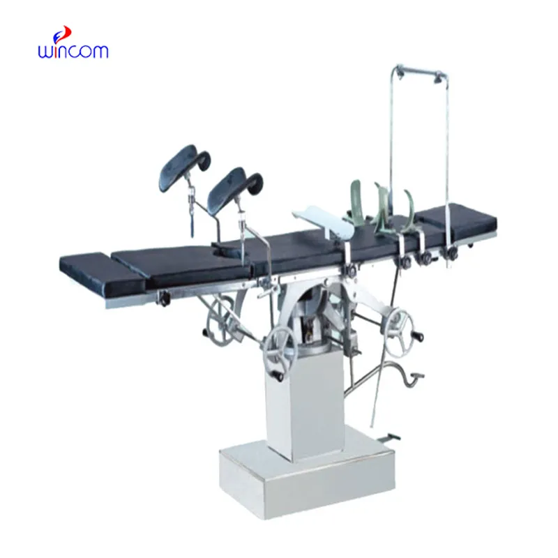
The delivery bed is well-designed and reliable. Our staff finds it simple to operate, and patients feel comfortable using it.
To protect the privacy of our buyers, only public service email domains like Gmail, Yahoo, and MSN will be displayed. Additionally, only a limited portion of the inquiry content will be shown.
Could you share the specifications and price for your hospital bed models? We’re looking for adjus...
We are planning to upgrade our imaging department and would like more information on your mri machin...
E-mail: [email protected]
Tel: +86-731-84176622
+86-731-84136655
Address: Rm.1507,Xinsancheng Plaza. No.58, Renmin Road(E),Changsha,Hunan,China

 af
af
 es
es
 ar
ar
 tr
tr
 sw
sw
 pt
pt
 th
th
 ur
ur
 bn
bn
 ne
ne
 vi
vi
 km
km
 lo
lo
 de
de
 ru
ru
 fi
fi
 nl
nl
 fa
fa
 fr
fr
 ko
ko