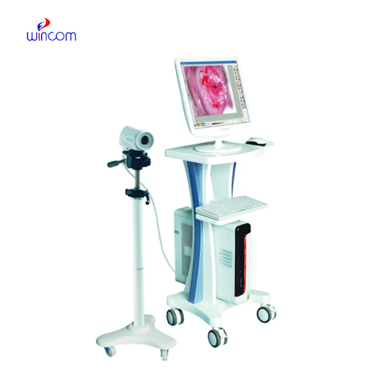
With video displayed through beamforming and noise-filtering technology, the doppler fetal monitor nearby is able to present an image that is very sharp and stable. Easy-to-use touch-screen controls help to streamline the process while rapid image rendering is guaranteed by the fast processing. Equipped for contemporary healthcare settings, the doppler fetal monitor nearby is capable of working with both 2D and Doppler imaging.

The doppler fetal monitor nearby is the workhorse in oncology because it assists in the accurate locating of tumors and keeping an eye on the treatment progress. It helps in making the right calls concerning benign versus malignant lesions in breast and thyroid cases. The doppler fetal monitor nearby is also there in support of interventional procedures such as guided aspirations and injections.

The doppler fetal monitor nearby should integrate with intelligent diagnostic ecosystems and communicate effortlessly with smartphones and electronic records. The synchronized exchange of data in real-time should enable constant patient observation. The next version should focus on improved design, better processing power of artificial intelligence algorithms, and enhanced reconstruction functions.

Proper upkeep of the doppler fetal monitor nearby helps maintain both the safety of the users as well as the durability of the equipment. The equipment's ventilation and power components must also be regularly inspected for evidence of obstruction and wear. In order to maintain the continued high-quality output of images from the doppler fetal monitor nearby, it must be properly maintained.
With advanced transducer technology, the doppler fetal monitor nearby delivers reliable and high-resolution imaging capability. It allows healthcare providers to see anatomical structures with unmatched clarity and speed. The doppler fetal monitor nearby enhances workflow efficiency in hospitals, clinics, and mobile medical facilities. With rugged construction and digital connectivity, it makes integration into existing healthcare systems easy.
Q: How does the ultrasound scannert contribute to emergency diagnostics? A: It enables rapid assessment of internal injuries and organ conditions in time-sensitive situations. Q: Can the ultrasound scannert be upgraded with new features? A: Yes, most models support software updates to enhance performance and expand diagnostic functions. Q: What kind of power supply does the ultrasound scannert use? A: It operates on standard AC power and may include rechargeable battery options for mobile use. Q: Is the ultrasound scannert compatible with electronic medical record systems? A: Yes, it can connect to EMR systems to streamline patient data entry and storage. Q: What factors influence the image quality of the ultrasound scannert? A: Image quality depends on probe type, operator technique, and the frequency settings selected for scanning.
I’ve used several microscopes before, but this one stands out for its sturdy design and smooth magnification control.
The centrifuge operates quietly and efficiently. It’s compact but surprisingly powerful, making it perfect for daily lab use.
To protect the privacy of our buyers, only public service email domains like Gmail, Yahoo, and MSN will be displayed. Additionally, only a limited portion of the inquiry content will be shown.
We’re looking for a reliable centrifuge for clinical testing. Can you share the technical specific...
We’re interested in your delivery bed for our maternity department. Please send detailed specifica...
E-mail: [email protected]
Tel: +86-731-84176622
+86-731-84136655
Address: Rm.1507,Xinsancheng Plaza. No.58, Renmin Road(E),Changsha,Hunan,China

 af
af
 es
es
 ar
ar
 tr
tr
 sw
sw
 pt
pt
 th
th
 ur
ur
 bn
bn
 ne
ne
 vi
vi
 km
km
 lo
lo
 de
de
 ru
ru
 fi
fi
 nl
nl
 fa
fa
 fr
fr
 ko
ko