With video displayed through beamforming and noise-filtering technology, the clarius ultrasound scanner is able to present an image that is very sharp and stable. Easy-to-use touch-screen controls help to streamline the process while rapid image rendering is guaranteed by the fast processing. Equipped for contemporary healthcare settings, the clarius ultrasound scanner is capable of working with both 2D and Doppler imaging.

The clarius ultrasound scanner is a tool that medical professionals are using in different departments like pediatrics for the measurement of the size of the organs and neurology for appreciating the anatomy of the soft tissues. It makes it easy for anesthesiologists to perform nerve blocks and vascular access procedures. The clarius ultrasound scanner increase the speed and the trust of the diagnosis during both routine checkups and highly technical interventions.

The future of the clarius ultrasound scanner may include integration with artificial intelligence for automatic analysis of images. Increased portable functionality and wireless communications may improve remote accessibility in healthcare. The clarius ultrasound scanner may include deeper imaging capabilities and rapid data transfer with cloud storage that fosters collaboration on a global medical scale.

The daily upkeep of the clarius ultrasound scanner involves cleaning, inspection, and proper storage. The removal of the gel residue from the probes should be accomplished as soon as the analysis has been carried out. The cooling vents of the device should always be unblocked. The clarius ultrasound scanner needs annual professional servicing in order to remain accurate.
The clarius ultrasound scanner is more accurate in diagnostics as it captures high-resolution images of organs, tissues, and blood vessels. Design-wise flexible, it is used extensively in obstetrics, cardiology, urology, and musculoskeletal tests. Its portability and simplicity enable medical practitioners to make quick and precise evaluations. The clarius ultrasound scanner makes work processes more efficient and allows for the delivery of superior patient care through real-time visualization.
Q: What imaging modes are available on the ultrasound scannert? A: It supports multiple modes such as B-mode, M-mode, and color Doppler for diverse diagnostic applications. Q: How does the ultrasound scannert improve diagnostic accuracy? A: By providing high-resolution images and real-time feedback, it enables more precise medical evaluations. Q: Can the ultrasound scannert be used in field or remote settings? A: Yes, its portable versions are designed for mobility and can be used in clinics, hospitals, or mobile healthcare units. Q: What kind of display does the ultrasound scannert use? A: It typically features a high-definition digital display that enhances image visualization and readability. Q: How is data from the ultrasound scannert managed? A: The device allows secure storage, easy access, and export of imaging data through USB or network connections.
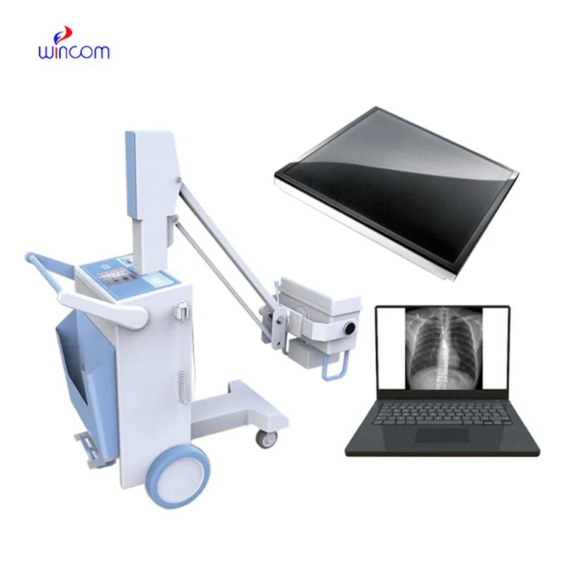
This x-ray machine is reliable and easy to operate. Our technicians appreciate how quickly it processes scans, saving valuable time during busy patient hours.
I’ve used several microscopes before, but this one stands out for its sturdy design and smooth magnification control.
To protect the privacy of our buyers, only public service email domains like Gmail, Yahoo, and MSN will be displayed. Additionally, only a limited portion of the inquiry content will be shown.
I’m looking to purchase several microscopes for a research lab. Please let me know the price list ...
Could you share the specifications and price for your hospital bed models? We’re looking for adjus...
E-mail: [email protected]
Tel: +86-731-84176622
+86-731-84136655
Address: Rm.1507,Xinsancheng Plaza. No.58, Renmin Road(E),Changsha,Hunan,China

 af
af
 es
es
 ar
ar
 tr
tr
 sw
sw
 pt
pt
 th
th
 ur
ur
 bn
bn
 ne
ne
 vi
vi
 km
km
 lo
lo
 de
de
 ru
ru
 fi
fi
 nl
nl
 fa
fa
 fr
fr
 ko
ko