The airport x ray machine images integrates the principles of ergonomics with high-quality imaging technologies. The easy-to-use system enables users to navigate with ease. The system also integrates automatic calibration functionality to ensure accurate outcomes. The airport x ray machine images supports digital storage and retrieval solutions that offer healthcare providers easy access to diagnostic images.

In operating rooms, the airport x ray machine images offers real-time imaging assistance in orthopedic and spinal surgeries. It helps surgeons confirm instrument positioning and bone alignment during surgery. The airport x ray machine images guarantees accuracy and dependability in real-time intraoperative decision-making.

The airport x ray machine images will move further forward with advances in detector materials and digital processing. Future systems will provide better image quality at much lower radiation doses. With more advanced AI-assisted workflows, the airport x ray machine images will enable radiologists to spend more time on clinical interpretation and less on hand-tweaking.

The airport x ray machine images requires careful attention to ensure imaging accuracy and equipment safety. The housekeeping staff must test system performance on a regular basis, including tube voltage, exposure timing, and detector status. The airport x ray machine images should always be turned off when being cleaned and checked routinely for mechanical or electrical wear.
The airport x ray machine images is intended to generate high-quality radiographic images that can capture internal details exceptionally. The device has the capability of being used in several medical functions such as trauma analysis and disease diagnosis. The portable and efficient airport x ray machine images helps in speeding up diagnosis and providing better treatment to the patient.
Q: What are the main components of an x-ray machine? A: The main components include the x-ray tube, control panel, collimator, image receptor, and protective housing, all working together to produce diagnostic images. Q: How should an x-ray machine be maintained? A: Regular inspection, calibration, and cleaning are essential to keep the x-ray machine operating accurately and safely over time. Q: What industries use x-ray machines besides healthcare? A: X-ray machines are also used in security screening, industrial testing, and materials inspection to identify defects or hidden items. Q: Why is calibration important for an x-ray machine? A: Calibration ensures that the machine delivers accurate radiation doses and consistent image quality, which is crucial for reliable diagnostics. Q: How long does an x-ray machine typically last? A: With proper maintenance, an x-ray machine can remain operational for over a decade, depending on usage frequency and environmental conditions.
The centrifuge operates quietly and efficiently. It’s compact but surprisingly powerful, making it perfect for daily lab use.
We’ve been using this mri machine for several months, and the image clarity is excellent. It’s reliable and easy for our team to operate.
To protect the privacy of our buyers, only public service email domains like Gmail, Yahoo, and MSN will be displayed. Additionally, only a limited portion of the inquiry content will be shown.
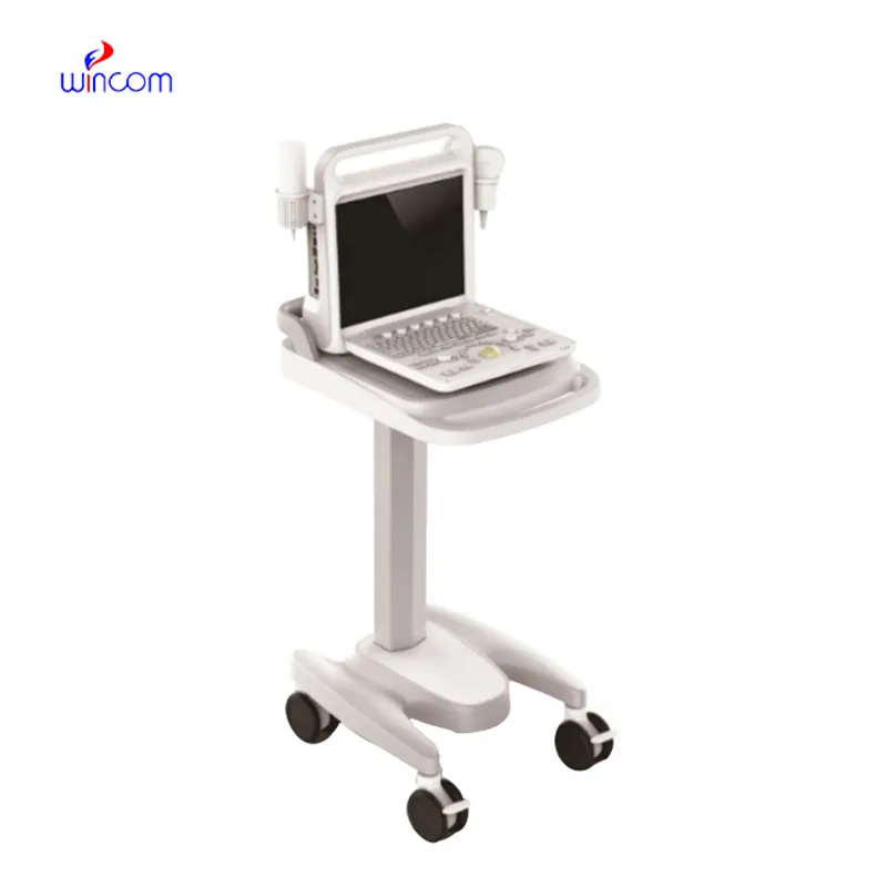
We’re currently sourcing an ultrasound scanner for hospital use. Please send product specification...
Hello, I’m interested in your water bath for laboratory applications. Can you confirm the temperat...
E-mail: [email protected]
Tel: +86-731-84176622
+86-731-84136655
Address: Rm.1507,Xinsancheng Plaza. No.58, Renmin Road(E),Changsha,Hunan,China

 af
af
 es
es
 ar
ar
 tr
tr
 sw
sw
 pt
pt
 th
th
 ur
ur
 bn
bn
 ne
ne
 vi
vi
 km
km
 lo
lo
 de
de
 ru
ru
 fi
fi
 nl
nl
 fa
fa
 fr
fr
 ko
ko